Safety

DIOR® Preclinical Program


Coronary arteries of 33 domestic pig were treated with the DIOR® drug-eluting balloon in a time-dependent matter. Arteries were dissected and sent to a blinded laboratory for paclitaxel determination. Histomorphometry and histopathology were performed two weeks after dilatation.
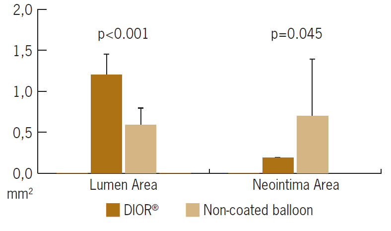
Lumen Area and Neointima area (mm²) at FU

1

Vessels treated with DIOR® DEB show significantly higher lumen area and significantly lower neointima area two weeks after dilatation compared to uncoated balloon treatment.

Area Dior


1

Pósa A., et al. Optimization of drug-eluting balloon use for safety and efficacy: Evaluation of the second generation paclitaxel-eluting DIOR-balloon in porcine coronary arteries. CCI 2010; 76(3):395-403


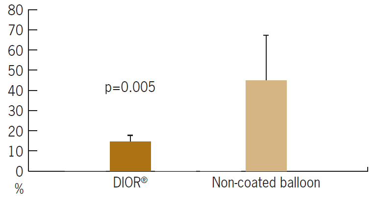
Area Stenosis (%) at FU

1

Vessels treated with DIOR® DEB show significantly lower area of stenosis after two weeks follow-up compared to uncoated balloon treatment.

Area Steonsis Dior


1

Pósa A., et al. Optimization of drug-eluting balloon use for safety and efficacy: Evaluation of the second generation paclitaxel-eluting DIOR-balloon in porcine coronary arteries. CCI 2010; 76(3):395-403


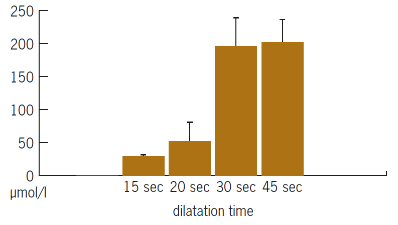
Tissue paclitaxel concentration (μmol/l)

1

Inflation of 45s with DIOR® DEB leads to long presence and high concentration of paclitaxel in the arterial wall – crucial for inhibition of neointimal proliferation and restenosis.

Paclitaxel Concentration Dior


1

Pósa A., et al. Optimization of drug-eluting balloon use for safety and efficacy: Evaluation of the second generation paclitaxel-eluting DIOR-balloon in porcine coronary arteries. CCI 2010; 76(3):395-403